Biochemistry · Proteins · Quiz

Protein structure levels quiz: primary to quaternary, with answers

Proteins fold in four levels: the amino acid sequence (primary), local backbone shapes (secondary), the full 3D fold of one chain (tertiary), and how separate chains fit together (quaternary). Exam questions almost always come down to one thing: which bonds hold each level together. Answer each question, then read the explanation, even when you get it right.

Learn it by doing: answer one practice problem and see why right away.

Try a practice problem now

On this pageThe quizSummary tableDenaturation practiceFAQ

The quiz: 12 questions by level

Pick an answer and press Check. A wrong pick tells you why that option is wrong, then explains the right one. Use Tab to move, the arrow keys to choose, and Enter or the Check button to check.

Primary structure

The amino acid sequence, joined by peptide bonds.

  1. Q1. Primary structure is held together by which bond?
    Answer and explanation for question 1

    Answer: B. Peptide bonds between amino acids. Primary structure is the order of amino acids, written from the N-terminus (free amino group) to the C-terminus (free carboxyl group). Each amino acid is joined to the next by a covalent peptide (amide) bond. Example: insulin’s A chain is 21 amino acids long and its B chain is 30.

    • A. Hydrogen bonds between backbone atoms: Not this one. Backbone H-bonds make secondary structure, not the sequence itself.
    • B. Peptide bonds between amino acids: Yes. The sequence is a chain of amino acids linked by covalent peptide bonds.
    • C. Disulfide bonds between cysteines: Not this one. Disulfides are covalent, but they link side chains during folding (tertiary or quaternary structure). They don’t define the sequence.
    • D. Ionic bonds between side chains: Not this one. Ionic interactions between R groups (salt bridges) are tertiary structure.
  2. Q2. Why can’t the backbone rotate freely around the peptide bond?
    Answer and explanation for question 2

    Answer: A. The peptide bond has partial double-bond character. Resonance spreads electrons across the C=O and C–N, so the C–N bond is partly double. That keeps the six atoms of the peptide group flat (planar), and the bond is almost always trans (bonds just before a proline are the main exception, and can be cis). The backbone bends at the two single bonds on either side of each alpha carbon, called phi (φ) and psi (ψ).

    • A. The peptide bond has partial double-bond character: Yes. Resonance gives the C–N bond partial double-bond character, so it doesn’t rotate.
    • B. The peptide bond is ionic: Not this one. The peptide bond is covalent, not ionic.
    • C. Side chains block it: Not this one. Bulky side chains limit some angles, but the peptide bond itself is stiff for a different reason.
    • D. It is held by a hydrogen bond: Not this one. H-bonds form between different peptide groups, not across the C–N bond itself.

Secondary structure

Local backbone shapes: alpha helices and beta sheets.

  1. Q3. Secondary structure (alpha helices and beta sheets) is stabilized mainly by:
    Answer and explanation for question 3

    Answer: B. Hydrogen bonds between backbone C=O and N–H groups. Secondary structure is the local shape of the backbone. The C=O of one peptide group hydrogen bonds to the N–H of another. R groups are not part of these H-bonds.

    • A. Hydrogen bonds between side chains: Not this one. Side-chain H-bonds help tertiary structure. Secondary structure uses backbone H-bonds; side chains still affect how readily a helix or sheet forms.
    • B. Hydrogen bonds between backbone C=O and N–H groups: Yes. The backbone carbonyl O of one peptide group bonds to the amide H of another.
    • C. Hydrophobic interactions: Not this one. The hydrophobic effect is the main driver of tertiary folding, not helices and sheets.
    • D. Disulfide bonds: Not this one. Disulfides are covalent links between cysteine side chains (tertiary or quaternary).
  2. Q4. In an alpha helix, the C=O of residue n hydrogen bonds to the N–H of which residue?
    Answer and explanation for question 4

    Answer: C. n + 4. The alpha helix is right-handed, with about 3.6 residues per turn. Each C=O bonds to the N–H four residues further along, so the H-bonds run roughly parallel to the helix axis. The side chains point outward, away from the axis.

    • A. n + 1: Not this one. Too close. The next residue is right beside it in the chain and can’t line up for an H-bond along the helix.
    • B. n + 2: Not this one. Still too close. Count one full turn of the helix (about 3.6 residues) and round up.
    • C. n + 4: Yes. Each C=O bonds to the N–H four residues further along.
    • D. A residue on a different strand: Not this one. H-bonds between separate strands describe a beta sheet, not a helix.
  3. Q5. Which amino acid is most likely to break an alpha helix?
    Answer and explanation for question 5

    Answer: C. Proline. Proline’s side chain loops back and bonds to its own backbone nitrogen. That leaves no N–H to donate an H-bond and locks the phi angle, so it usually disrupts the regular helix pattern. Glycine also weakens helices, for the opposite reason: it is so flexible that it rarely holds the helix shape.

    • A. Alanine: Not this one. Alanine is one of the strongest helix formers.
    • B. Leucine: Not this one. Leucine is a strong helix former too.
    • C. Proline: Yes. Proline is the classic helix breaker.
    • D. Glutamate: Not this one. Glutamate is fine in helices. It’s actually a common helix former.
  4. Q6. Which statement about beta sheets is TRUE?
    Answer and explanation for question 6

    Answer: B. Strands can run parallel or antiparallel, with H-bonds between neighboring strands. Beta strands are stretched-out segments of backbone lined up side by side. Their backbone C=O and N–H groups hydrogen bond across strands, and the side chains stick out above and below the pleats. Antiparallel sheets have straighter, slightly more stable H-bonds than parallel sheets.

    • A. H-bonds form within a single strand, as in a helix: Not this one. That’s the helix pattern. Sheets bond strand to strand.
    • B. Strands can run parallel or antiparallel, with H-bonds between neighboring strands: Yes. Both kinds exist, and the H-bonds always link one strand to the next.
    • C. Beta sheets are held together by disulfide bonds: Not this one. Sheets use backbone H-bonds. Disulfides are a side-chain link.
    • D. Side chains all point to the same side of the sheet: Not this one. Side chains alternate, above and below the sheet.

Tertiary structure

The full 3D fold of one chain, held by side-chain (R-group) interactions.

  1. Q7. What is the main driving force for a soluble protein folding into its tertiary structure?
    Answer and explanation for question 7

    Answer: B. The hydrophobic effect: nonpolar side chains get buried away from water. Burying nonpolar side chains (Leu, Ile, Val, Phe and others) in the core frees the water molecules that were ordered around them. Soluble proteins tend to bury many nonpolar side chains and expose many polar or charged ones, though there are exceptions. Which side chains are nonpolar? See the amino acid quiz.

    • A. Disulfide bond formation: Not this one. Disulfides lock a fold in place, but many proteins have none at all.
    • B. The hydrophobic effect: nonpolar side chains get buried away from water: Yes. Burying nonpolar side chains is the biggest single push toward the folded shape.
    • C. Peptide bond formation: Not this one. Peptide bonds are made on the ribosome before folding. That’s primary structure.
    • D. Ionic bonds between the N- and C-termini: Not this one. Salt bridges contribute, but they’re a small part of folding.
  2. Q8. Which interaction is NOT a side-chain (R-group) interaction in tertiary structure?
    Answer and explanation for question 8

    Answer: D. An H-bond between backbone C=O and N–H in a helix. Tertiary structure is held by R-group interactions: hydrophobic packing, ionic bonds (salt bridges), side-chain H-bonds, van der Waals contacts, and covalent disulfide bonds. Backbone-to-backbone H-bonds are secondary structure. Whether a Lys or Asp side chain is charged depends on pH; practice that on the isoelectric point page.

    • A. A salt bridge between Lys and Asp: Not this one. Lys (+) and Asp (−) side chains attract each other. That ionic interaction counts as tertiary.
    • B. A disulfide bond between two Cys: Not this one. A disulfide links two cysteine side chains, so it’s tertiary (or quaternary, between chains).
    • C. An H-bond between Ser and Gln side chains: Not this one. Polar side chains hydrogen bonding with each other is a tertiary interaction.
    • D. An H-bond between backbone C=O and N–H in a helix: Yes. That one is backbone to backbone, so it’s secondary structure.
  3. Q9. A disulfide bond is:
    Answer and explanation for question 9

    Answer: B. A covalent S–S bond formed by oxidizing two cysteine thiols. Two cysteine –SH groups are oxidized to make –S–S– (the linked pair is called cystine). Disulfides are common in secreted proteins such as antibodies and insulin, and rare inside the cytosol, where conditions are reducing.

    • A. A noncovalent bond between two methionines: Not this one. Methionine’s sulfur is a thioether (no –SH), so it can’t form disulfides.
    • B. A covalent S–S bond formed by oxidizing two cysteine thiols: Yes. Two –SH groups are oxidized to one –S–S– link.
    • C. A hydrogen bond between two cysteines: Not this one. It’s covalent, which makes it much stronger than an H-bond.
    • D. Broken by urea alone: Not this one. Urea disrupts noncovalent interactions. To break a disulfide you need a reducing agent such as β-mercaptoethanol or DTT.

Quaternary structure

Two or more separate chains (subunits) packed together.

  1. Q10. Which protein has quaternary structure?
    Answer and explanation for question 10

    Answer: B. Hemoglobin. Hemoglobin is a tetramer of two α and two β subunits (α2β2), each carrying its own heme. Its subunits are held together by noncovalent interactions and affect each other, which is why its oxygen binding is cooperative (a sigmoidal curve). Myoglobin is one chain with one heme and a hyperbolic curve.

    • A. Myoglobin: Not this one. Myoglobin is a single chain, so it only goes up to tertiary structure.
    • B. Hemoglobin: Yes. Hemoglobin has four subunits.
    • C. A single alpha helix: Not this one. A helix is secondary structure within one chain.
    • D. Any protein with disulfide bonds: Not this one. Disulfides can sit within one chain. Quaternary structure needs two or more separate chains.
  2. Q11. Which forces can hold subunits together in quaternary structure?
    Answer and explanation for question 11

    Answer: C. The same forces as tertiary: hydrophobic, ionic, H-bonds, and sometimes disulfides. Quaternary structure uses the same toolkit as tertiary structure, applied between chains instead of within one. Mostly that means weak noncovalent contacts; some proteins, such as antibodies, add disulfide bonds between chains.

    • A. Only peptide bonds: Not this one. If subunits were joined by peptide bonds, they would be one chain.
    • B. Only disulfide bonds: Not this one. Some proteins use disulfides between chains (antibodies, and insulin’s A and B chains), but many, like hemoglobin, have none between subunits.
    • C. The same forces as tertiary: hydrophobic, ionic, H-bonds, and sometimes disulfides: Yes. Same toolkit, applied between chains.
    • D. Only backbone H-bonds: Not this one. Subunit contacts mostly involve side chains.

Denaturation

What survives when a protein unfolds?

  1. Q12. A protein is heated until it unfolds (denatures). Which level of structure survives?
    Answer and explanation for question 12

    Answer: A. Primary. Denaturation disrupts the noncovalent interactions (H-bonds, hydrophobic, ionic) that hold secondary, tertiary and quaternary structure. Peptide bonds are not broken, so the sequence is intact. Breaking peptide bonds takes hydrolysis (proteases, or strong acid with prolonged heating), and that’s a different process.

    • A. Primary: Yes. Heat doesn’t cut peptide bonds, so the sequence stays intact.
    • B. Secondary: Not this one. Heat disrupts backbone H-bonds, so helices and sheets can fall apart.
    • C. Tertiary: Not this one. Heat disrupts noncovalent side-chain interactions, so the 3D fold is lost. That’s what denaturing means.
    • D. Quaternary: Not this one. Most subunit contacts are noncovalent and can be disrupted by heat; some proteins also have interchain disulfides.

Want more like this? Quiz yourself on protein structure, amino acids and enzymes, with questions that come back when you miss them.

Try 3 days free

3-day free trial, no card needed.

Summary table: what holds it together, what breaks it

The four levels of protein structure
LevelWhat it isHeld together byExampleSurvives denaturation?
PrimaryAmino acid sequence (N → C)Covalent peptide bondsThe 21-residue sequence of insulin’s A chainYes. Only hydrolysis (proteases, strong acid) cuts peptide bonds
SecondaryLocal backbone shapesH-bonds between backbone C=O and N–HAlpha helix (3.6 residues per turn, n → n + 4); beta sheetNo
TertiaryFull 3D fold of one chainR-group interactions: hydrophobic effect, ionic (salt bridges), side-chain H-bonds, van der Waals, disulfides (covalent)MyoglobinNo. Disulfides stay unless a reducing agent is added
QuaternaryTwo or more chains packed togetherSame as tertiary, between chainsHemoglobin (α2β2)No
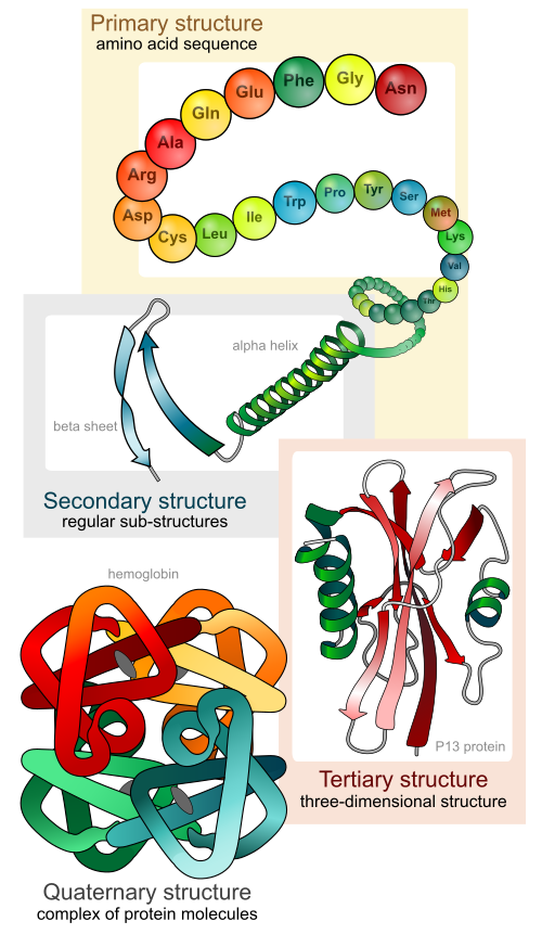
Diagram of the four levels of protein structure: a chain of amino acids (primary), an alpha helix and a beta pleated sheet (secondary), a folded single chain (tertiary), and several folded chains packed together, shown as hemoglobin (quaternary).
The four levels. Image: LadyofHats (Mariana Ruiz Villarreal), Wikimedia Commons, public domain.

Quick denaturation practice

For each agent, pick the main thing it disrupts. One best answer each.

What does each agent disrupt?
Show the practice answers
Denaturation practice answers
AgentDisruptsWhy
HeatH-bonds and hydrophobic interactionsHeat adds energy that shakes apart weak noncovalent interactions. It doesn’t cut peptide bonds.
Urea (8 M)H-bonds and hydrophobic interactionsUrea competes for H-bonds and helps nonpolar groups dissolve in water. It leaves covalent bonds alone.
Extreme pHIonic interactions (salt bridges)Changing the pH changes which side chains carry a charge, so salt bridges break.
β-mercaptoethanol or DTTDisulfide bondsThese are reducing agents: they turn –S–S– back into two –SH groups.
SDS (a detergent)H-bonds and hydrophobic interactionsSDS coats the nonpolar regions and unfolds the chain. That’s how SDS-PAGE works; a reducing agent is often added too, to cut disulfides.
Trypsin (a protease)Peptide bondsProteases hydrolyze peptide bonds. That’s digestion, not denaturation.

Anfinsen’s experiment. Ribonuclease A was unfolded with 8 M urea plus β-mercaptoethanol, which also reduced its four disulfide bonds. Removing β-mercaptoethanol before urea let the wrong disulfide pairs form, so little activity returned. Adding a small amount of β-mercaptoethanol let the bonds reshuffle and restored activity. The sequence carried the information needed for the native fold.

FAQ

What are the four levels of protein structure?

Primary (the amino acid sequence), secondary (alpha helices and beta sheets formed by backbone H-bonds), tertiary (the 3D fold of one chain, held by side-chain interactions), and quaternary (two or more chains assembled together, such as hemoglobin).

What’s the difference between secondary and tertiary structure?

Secondary structure comes from hydrogen bonds between backbone atoms only. Tertiary structure comes from interactions between side chains (R groups): hydrophobic, ionic, hydrogen bonds and disulfide bonds.

Does denaturation break peptide bonds?

No. Denaturation disrupts the noncovalent interactions of secondary, tertiary and quaternary structure, so the primary sequence stays intact. Breaking peptide bonds requires hydrolysis.

Does every protein have quaternary structure?

No. Only proteins made of two or more polypeptide chains do. Myoglobin is a single chain, so it stops at tertiary structure.

Sources

Ready to lock in protein structure before your exam? Keep practicing with instant feedback.

Try 3 days free

3-day free trial, no card needed.

Ready to practice? Answer a problem and get instant feedback on every step.

Try a practice problem now